


THANK YOU FOR SUBSCRIBING
Weekly Brief
Be first to read the latest tech news, Industry Leader's Insights, and CIO interviews of medium and large enterprises exclusively from Healthcare Tech Outlook
A featured contribution from Leadership Perspectives: a curated forum reserved for leaders nominated by our subscribers and vetted by our Healthcare Tech Outlook Advisory Board.

M.D.
Emerging Strategies in Autologous Breast Reconstruction
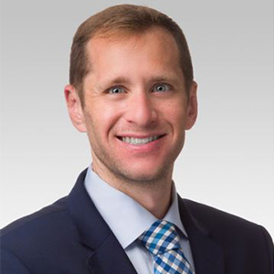
Chad M. Teven
Microsurgery and Complex Reconstruction
Division of Plastic Surgery
Northwestern University Feinberg School of Medicine
Breast cancer is the most common non-skin malignancy in women in the United States, with nearly 300,000 new cancers diagnosed per year, as reported by the Centers for Disease Control and Prevention.1As the number of breast cancer cases continues to rise, so too do the number of breast reconstruction procedures. This upward trend is the result of increased awareness, improved reconstructive techniques and options, and shifting mastectomy patterns.2 In particular, more women with unilateral breast cancer are opting for bilateral mastectomy -including contralateral prophylactic mastectomy – instead of unilateral mastectomy or breast conservation. In recent years, several authors have compared autologous and prosthetic reconstruction.3 Although there is significant emerging data to support improved long-term quality of life and patient satisfaction after autologous reconstruction, rates of autologous reconstruction have remained relatively stagnant in the last two decades.4 This may be explained by reduced reimbursement and increased technical complexity associated with autologous reconstruction relative to prosthetic reimbursement. We believe, however, that autologous reconstruction is superior and have thus adopted strategies in our practice to support offering an increasing number of patients the option of autologous reconstruction.
Early descriptions of breast reconstruction using abdominally-based tissue included skin, subcutaneous (i.e., adipose) tissue, muscle, and fascia from the abdominal wall.5This resulted in significant morbidity at the donor site, including high rates of hernia, bulge, and abdominal weakness. More recently, muscle- and fascia-sparing techniques, including the deep inferior epigastric artery perforator (DIEP) flap and superficial inferior epigastric artery (SIEA) flap, have been employed to reduce donor site morbidity. These techniques require increased time and skill during the harvest of the flap. Our practice employs the use of these and other perforator flaps (i.e., muscle-sparing) in nearly every autologous breast reconstruction performed, thus improving clinical outcomes in our patient population. A clear understanding of perforator anatomy, technical experience and expertise, and the use of CT-guided imaging to identify blood vessels of interest preoperatively - and thus direct surgical dissection – are critical factors in the optimization of these techniques.6
A second important strategy to improve outcomes in breast reconstruction is the use of enhanced recovery after surgery (ERAS) techniques.7 ERAS denotes a perioperative approach that consists of multidisciplinary, multimodal, and evidence-based strategies aimed at improving recovery after surgery, including reduced opioid use and hospital length of stay. At our institution, strong collaboration among the surgery and anesthesia teams is responsible for the seamless implementation andsuccess of ERAS. This, in conjunction, with improved operative technique, has resulted in reduced operative times, reduced time to discharge after surgery, and improved pain control.
 Michael A. Howard, M.D.
A further advance in recent years that is in part responsible for the success of autologous breast reconstruction at our and other centers is the improved ability to monitor free flaps post-operatively. One concerning complication with free tissue transfer is the interruption of adequate perfusion to the tissue, often due to arterial thrombosis or venous congestion. In such cases, early identification of a vascular issue is paramount to tissue salvage. Many methods for flap monitoring exists, each with its own advantages and disadvantages.8 Traditionally, monitoring of flap perfusion is based on the clinical examination, which includes the appearance of the flap as well as the presence or absence of audible measures of arterial and venous flow.Our center employs the T-Stat 2 Tissue Oximeter (Spectros Medical Devices, Inc., Houston, TX), which uses visible light spectroscopy to evaluate both arterial and venous perfusion. Specific advantages of this device include rapid identification of flap compromise – often before a clinical change can be appreciated on exam – and the ability to remotely monitor the flap (i.e., information regarding flap perfusion is transmitted in real-time to the surgical team both inside and out of the hospital). This further allows for the mitigation of monitoring risks associated with clinical evaluation at the bedside (i.e., often by nursing and trainees), including limited experience potentially resulting in a reduction of correct interpretation, the need for frequent uncovering and palpation of the target site (i.e., potentially leading to ischemia- or trauma-induced vasoconstriction), and limited personnel with staff shortages.
As the incidence of breast cancer increases, alongside continued support from emerging data on the use of autologous tissue in breast reconstruction, reconstructive surgeons ought to embrace autologous-based techniques for reconstruction. Indeed, by adopting and implementing strategies that are borne from new ideas and technology, surgeons will be better suited to perform autologous breast reconstruction safely and efficiently. Furthermore, by reducing complications, surgical duration, and hospital length of stay, issues regarding cost and reimbursement discrepancies between prosthetic and autologous reconstruction will narrow. As demonstrated in our own practice, we believe that significant advances in techniques and associated technology in breast reconstruction will result in optimized patient outcomes.
References
1. United States Cancer Statistics: Data Visualizations. Center for Disease Control and Prevention. 2022. Available from: https://gis.cdc.gov/Cancer/USCS/#/Trends/. Accessed September 11, 2022.
2. Panchal H, Matros E. Current trends in post-mastectomy breast reconstruction. Plast Reconstr Surg. 2017;140(5):7S-13S.
3. Santosa KB, Qi J, Kim HM, Hamill JB, Wilkins EG, Pusic AL. Long-term patient-reported outcomes in postmastectomy breast reconstruction. JAMA Surg. 2018;153(10):891-899.
4. Yueh JH, Slavin SA, Adesiyun T, et al. Patient satisfaction in postmastectomy breast reconstruction: A comparative evaluation of DIEP, TRAM, latissimus flap, and implant techniques. Plast Reconstr Surg. 2010;125(6):1585-1595.
5. Uroskie TW, Colen LB. History of breast reconstruction. Semin Plast Surg. 2004;18(2):65-69.
6. O’Connor EF, Rozen WM, Chowdhry M, et al. Preoperative computed tomography angiography for planning DIEP flap breast reconstruction reduces operative time and overall complications. Gland Surg. 2016;5(2):93-98.
7. Hammond JB, Thomas O, Jogerst K, et al. Same-day discharge is safe and effective after implant-based breast reconstruction. Ann Plast Surg. 2020; 87(2):144-149.
8. Teven CM, Ooi ASH, Inbal A, Chang DW. Implantable Doppler monitoring of buried free flaps during vascularized lymph node transfer. J Surg Oncol 2017;116(3):371-377.
The articles from these contributors are based on their personal expertise and viewpoints, and do not necessarily reflect the opinions of their employers or affiliated organizations.
